Mata Chanan Devi Hospital started MDH INSTITUTE OF NEUROSCIENCES & RESEARCH for providing neurological and neurosurgical cover to residents of Janakpuri, Vikaspuri and Dwarka particularly and for any other region residents generally wef 1 Jan 2007 in a new and modern block of hospital. There is provision for evaluation and management of neurological / neurosurgical conditions, both for adults and children in the highest standards acceptable in any private sector. The institute has installed the most modern diagnostic equipments for diagnosis and management of Neurological / Neurosurgical diseases.
The department is well equipped to manage all types of neurological problems like strokes, epilepsy, infections of central nerves system (Meningitis, Encephalitis), Immunological (Multiple Sclerosis, AIDP, CIDP, Myasthenia Gravis) and degenerative (Parkinson Disease, Dementia including Alzheimer’s disease) disorders of CNS, peripheral nerve disorders (Neuropathies) and muscles disorders (Myopathies).
Facilities available include:
I) NICU / HDU
There is dedicated Neuro intensive care unit (NICU) ,18 Bedded and High Dependency Unit (HDU),10 Bedded, to cater for emergency, seriously ill neurology cases. The units are equipped with the latest monitoring devices and advanced life supporting systems needed for management of serious and complicated neurological / neurosurgical problems effectively with a patient / nurse ratio of 1:1. All related specialists for management of such cases like Cardiologist, Respiratory, Physicians, Nephrologists, Anaesthetists and Internists are available round the clock.
II) Stroke Unit
There is a dedicated Stroke unit to attend to cases of cerebovascular accidents / diseases. Consultant neurologists and an Endovascular Neurosurgeon are available for early management of all types of strokes. MRI with diffusion weighted and FLAIR images is used to diagnose acute cases within first 3 hrs of onset so that thrombolysis therapy can be effectively administered.
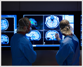
III) Diagnostic Facilities
Neuroimaging
- MRI: Latest MRI capable of doing MRI, MRA, and Diffusion weighted imaging is available. This can be used to obtain / spinal cord images, MR angiography, MR venography. Infarcts of the brain (Stroke cases) can be diagnosed in the hyper acute stage helping in decision on thrombolytic therapy. MRI spine imaging gives clarity about cases of spinal cord diseases requiring medical (Myelopathy) / surgical therapy (Tumors / IV disc diseases / fractures).
- Computerised Tomography (CT) : The latest state of the art multislice CT Scan, capable of acquiring images rapidly is available to investigate cranial / spinal
neurological conditions. This latest machine is capable of performing CT angiography (cranial / spinal) with precision as good as conventional angiography or DSA. It can provide 3D images useful for reconstructive surgery.
-
Transcranial Doppler(TCD) : This procedure is useful for diagnosis of vasospasm in cases of subarachnoid haemorrage (SAH). In cases of Transient Ischaemic Attack (TIA) territories of hit can be identified. TCD monitoring can also be done intraoperatively.
- Duplex Carotid Ultrasonography (Carotid Doppler) to study the carotid arteries flow study pattern in cerebrovascular disorders (stroke) is available.
- Digital Subtraction Angiography: The state of the art GE DSA machine is available for performing cerebral angiographies, carotid angioplasty, stenting aneurysm coiling and embolisation.
- All routine Digital X-rays and Ultrasound investigations are available and are routinely used.
Electrophysiology Lab:
- Electrophysiology (EEG) : Facilities to perform routine digital EEG, with the most modern equipment is available to diagnose cases of seizure, stroke etc.
- In seriously ill patients who can not be moved to the lab, bedside digital EEG can be performed.
To diagnose better seizure / epilepsy cases, Video EEG is available by which various types of seizure / behavioral abnormalities can be correlated with EEG.
- Nerve conduction studies: Portable Digital Myto Electromyographic machine is available. Motor and sensory nerve conduction studies can be done in polyneuropathy / traumatic (Plexus Injury) cases.
- Electromyography : can be done to identify neuropathy / myopathic diseases. Repetitive stimulation studies can be done to diagnosis Myasthenia Gravis.
- Evoked Potential Studies : Facilities to conduct Somatosensory Evoked Potentials (SSEP), Visual Evoked Potentials (VEP), Brainstem Auditory Evoked Potentials (BAEP) are available to diagnose spinal cord, visual pathways, brain stem pathways disorder.
 + (91) - (11) - 45582000/10